Uploads by Was a bee
Jump to navigation
Jump to search

For Was a bee (talk · contributions · Move log · block log · uploads · Abuse filter log)
This special page shows all uploaded files that have not been deleted; for those see the upload log.
| Date | Name | Thumbnail | Size | Description |
|---|---|---|---|---|
| 12:47, 18 June 2021 | Tractography - Parietopontine tract - animation 0.gif (file) |  |
22.39 MB | c:User:Rillke/bigChunkedUpload.js: == {{int:filedesc}} == {{Information |Description={{en|1=Tractography of the parietopontine tract. Animation. 3D Tractography data (by Fang-Cheng Yeh et al.) was superimposed on the skull and the brain from the BodyParts3D, a free 3D human body parts library. Superimposition was done by the Blender, a free and open-source 3D computer graphics software (file conversion method). Rendering was do... |
| 08:45, 6 June 2021 | Tractography - Parietopontine tract - animation 2.gif (file) |  |
10.73 MB | == {{int:filedesc}} == {{Information |Description={{en|1=Tractography of the parietopontine tract. Animation. 3D Tractography data was rendered by the Blender, a free and open-source 3D computer graphics software (file conversion method). Rendering was done by the SheepIt, a free distributed renderfarm for the Blender. {{Legend|Yellow|Yellow: Left parietopontine tract}} {{Legend|Red|Red: Right parietopontine tract}} }} |Source=T... |
| 07:51, 6 June 2021 | Tractography - Parietopontine tract - inferior view.png (file) |  |
997 KB | == {{int:filedesc}} == {{Information |Description={{en|1=Tractography of the parietopontine tract. 3D Tractography data (by Fang-Cheng Yeh et al.) was superimposed on the skull and the brain from the BodyParts3D, a free 3D human body parts library. Superimposition was done by the Blender, a free and open-source 3D computer graphics software (file conversion method). Rendering was done by the SheepIt, a free distributed renderfarm f... |
| 07:49, 6 June 2021 | Tractography - Parietopontine tract - superior view.png (file) |  |
996 KB | == {{int:filedesc}} == {{Information |Description={{en|1=Tractography of the parietopontine tract. 3D Tractography data (by Fang-Cheng Yeh et al.) was superimposed on the skull and the brain from the BodyParts3D, a free 3D human body parts library. Superimposition was done by the Blender, a free and open-source 3D computer graphics software (file conversion method). Rendering was done by the SheepIt, a free distributed renderfarm f... |
| 07:48, 6 June 2021 | Tractography - Parietopontine tract - lateral view (left).png (file) | .png) |
1.18 MB | == {{int:filedesc}} == {{Information |Description={{en|1=Tractography of the parietopontine tract. 3D Tractography data (by Fang-Cheng Yeh et al.) was superimposed on the skull and the brain from the BodyParts3D, a free 3D human body parts library. Superimposition was done by the Blender, a free and open-source 3D computer graphics software (file conversion method). Rendering was done by the SheepIt, a free distributed renderfarm f... |
| 07:47, 6 June 2021 | Tractography - Parietopontine tract - posterior view.png (file) |  |
980 KB | == {{int:filedesc}} == {{Information |Description={{en|1=Tractography of the parietopontine tract. 3D Tractography data (by Fang-Cheng Yeh et al.) was superimposed on the skull and the brain from the BodyParts3D, a free 3D human body parts library. Superimposition was done by the Blender, a free and open-source 3D computer graphics software (file conversion method). Rendering was done by the SheepIt, a free distributed renderfarm f... |
| 07:47, 6 June 2021 | Tractography - Parietopontine tract - lateral view (right).png (file) | .png) |
1.16 MB | == {{int:filedesc}} == {{Information |Description={{en|1=Tractography of the parietopontine tract. 3D Tractography data (by Fang-Cheng Yeh et al.) was superimposed on the skull and the brain from the BodyParts3D, a free 3D human body parts library. Superimposition was done by the Blender, a free and open-source 3D computer graphics software (file conversion method). Rendering was done by the SheepIt, a free distributed renderfarm f... |
| 07:46, 6 June 2021 | Tractography - Parietopontine tract - anterior view.png (file) |  |
996 KB | == {{int:filedesc}} == {{Information |Description={{en|1=Tractography of the parietopontine tract. 3D Tractography data (by Fang-Cheng Yeh et al.) was superimposed on the skull and the brain from the BodyParts3D, a free 3D human body parts library. Superimposition was done by the Blender, a free and open-source 3D computer graphics software (file conversion method). Rendering was done by the SheepIt, a free distributed renderfarm f... |
| 07:32, 6 June 2021 | Tractography - Parietopontine tract - animation 1.gif (file) |  |
14.73 MB | == {{int:filedesc}} == {{Information |Description={{en|1=Tractography of the parietopontine tract. Animation. 3D Tractography data (by Fang-Cheng Yeh et al.) was superimposed on the skull and the brain from the BodyParts3D, a free 3D human body parts library. Superimposition was done by the Blender, a free and open-source 3D computer graphics software (file conversion method). Rendering was done by the SheepIt, a free distributed r... |
| 02:11, 5 June 2021 | Tractography - Occipitopontine tract - animation 2.gif (file) |  |
10.96 MB | == {{int:filedesc}} == {{Information |Description={{en|1=Tractography of the occipitopontine tract. Animation. 3D Tractography data (by Fang-Cheng Yeh et al.) was rendered by the Blender, a free and open-source 3D computer graphics software (file conversion method). Rendering was done by the SheepIt, a free distributed renderfarm for the Blender. {{Legend|Yellow|Yellow: Left occipitopontine tract}} {{Legend|Red|Red: Right occipi... |
| 20:57, 4 June 2021 | Tractography - Occipitopontine tract - inferior view.png (file) |  |
1,021 KB | == {{int:filedesc}} == {{Information |Description={{en|1=Tractography of the occipitopontine tract. 3D Tractography data (by Fang-Cheng Yeh et al.) was superimposed on the skull and the brain from the BodyParts3D, a free 3D human body parts library. Superimposition was done by the Blender, a free and open-source 3D computer graphics software (file conversion method). Rendering was done by the SheepIt, a free distributed renderfarm... |
| 20:57, 4 June 2021 | Tractography - Occipitopontine tract - superior view.png (file) |  |
1,015 KB | == {{int:filedesc}} == {{Information |Description={{en|1=Tractography of the occipitopontine tract. 3D Tractography data (by Fang-Cheng Yeh et al.) was superimposed on the skull and the brain from the BodyParts3D, a free 3D human body parts library. Superimposition was done by the Blender, a free and open-source 3D computer graphics software (file conversion method). Rendering was done by the SheepIt, a free distributed renderfarm... |
| 20:56, 4 June 2021 | Tractography - Occipitopontine tract - posterior view.png (file) |  |
990 KB | == {{int:filedesc}} == {{Information |Description={{en|1=Tractography of the occipitopontine tract. 3D Tractography data (by Fang-Cheng Yeh et al.) was superimposed on the skull and the brain from the BodyParts3D, a free 3D human body parts library. Superimposition was done by the Blender, a free and open-source 3D computer graphics software (file conversion method). Rendering was done by the SheepIt, a free distributed renderfarm... |
| 20:56, 4 June 2021 | Tractography - Occipitopontine tract - lateral view (left).png (file) | .png) |
1.17 MB | == {{int:filedesc}} == {{Information |Description={{en|1=Tractography of the occipitopontine tract. 3D Tractography data (by Fang-Cheng Yeh et al.) was superimposed on the skull and the brain from the BodyParts3D, a free 3D human body parts library. Superimposition was done by the Blender, a free and open-source 3D computer graphics software (file conversion method). Rendering was done by the SheepIt, a free distributed renderfarm... |
| 20:53, 4 June 2021 | Tractography - Occipitopontine tract - lateral view (right).png (file) | .png) |
1.16 MB | == {{int:filedesc}} == {{Information |Description={{en|1=Tractography of the occipitopontine tract. 3D Tractography data (by Fang-Cheng Yeh et al.) was superimposed on the skull and the brain from the BodyParts3D, a free 3D human body parts library. Superimposition was done by the Blender, a free and open-source 3D computer graphics software (file conversion method). Rendering was done by the SheepIt, a free distributed renderfarm... |
| 20:52, 4 June 2021 | Tractography - Occipitopontine tract - anterior view.png (file) |  |
1,005 KB | == {{int:filedesc}} == {{Information |Description={{en|1=Tractography of the occipitopontine tract. 3D Tractography data (by Fang-Cheng Yeh et al.) was superimposed on the skull and the brain from the BodyParts3D, a free 3D human body parts library. Superimposition was done by the Blender, a free and open-source 3D computer graphics software (file conversion method). Rendering was done by the SheepIt, a free distributed renderfarm... |
| 20:13, 4 June 2021 | Tractography - Occipitopontine tract - animation 1.gif (file) |  |
14.27 MB | == {{int:filedesc}} == {{Information |Description={{en|1=Tractography of the occipitopontine tract. Animation. 3D Tractography data (by Fang-Cheng Yeh et al.) was superimposed on the skull and the brain from the BodyParts3D, a free 3D human body parts library. Superimposition was done by the Blender, a free and open-source 3D computer graphics software (file conversion method). Rendering was done by the SheepIt, a free distributed... |
| 20:32, 26 May 2021 | Tractography - Frontopontine tract - inferior view.png (file) |  |
635 KB | == {{int:filedesc}} == {{Information |Description={{en|1=Tractography of the frontopontine tract. 3D Tractography data (by Fang-Cheng Yeh et al.) was superimposed on the skull and the brain from the BodyParts3D, a free 3D human body parts library. Superimposition was done by the Blender, a free and open-source 3D computer graphics software (file conversion method). Rendering was done by the SheepIt, a free distributed renderfarm for... |
| 20:28, 26 May 2021 | Tractography - Frontopontine tract - superior view.png (file) |  |
633 KB | == {{int:filedesc}} == {{Information |Description={{en|1=Tractography of the frontopontine tract. 3D Tractography data (by Fang-Cheng Yeh et al.) was superimposed on the skull and the brain from the BodyParts3D, a free 3D human body parts library. Superimposition was done by the Blender, a free and open-source 3D computer graphics software (file conversion method). Rendering was done by the SheepIt, a free distributed renderfarm for... |
| 20:28, 26 May 2021 | Tractography - Frontopontine tract - posterior view.png (file) |  |
616 KB | == {{int:filedesc}} == {{Information |Description={{en|1=Tractography of the frontopontine tract. 3D Tractography data (by Fang-Cheng Yeh et al.) was superimposed on the skull and the brain from the BodyParts3D, a free 3D human body parts library. Superimposition was done by the Blender, a free and open-source 3D computer graphics software (file conversion method). Rendering was done by the SheepIt, a free distributed renderfarm for... |
| 20:28, 26 May 2021 | Tractography - Frontopontine tract - lateral view (left).png (file) | .png) |
742 KB | == {{int:filedesc}} == {{Information |Description={{en|1=Tractography of the frontopontine tract. 3D Tractography data (by Fang-Cheng Yeh et al.) was superimposed on the skull and the brain from the BodyParts3D, a free 3D human body parts library. Superimposition was done by the Blender, a free and open-source 3D computer graphics software (file conversion method). Rendering was done by the SheepIt, a free distributed renderfarm for... |
| 20:27, 26 May 2021 | Tractography - Frontopontine tract - lateral view (right).png (file) | .png) |
722 KB | == {{int:filedesc}} == {{Information |Description={{en|1=Tractography of the frontopontine tract. 3D Tractography data (by Fang-Cheng Yeh et al.) was superimposed on the skull and the brain from the BodyParts3D, a free 3D human body parts library. Superimposition was done by the Blender, a free and open-source 3D computer graphics software (file conversion method). Rendering was done by the SheepIt, a free distributed renderfarm for... |
| 20:27, 26 May 2021 | Tractography - Frontopontine tract - anterior view.png (file) |  |
626 KB | == {{int:filedesc}} == {{Information |Description={{en|1=Tractography of the frontopontine tract. 3D Tractography data (by Fang-Cheng Yeh et al.) was superimposed on the skull and the brain from the BodyParts3D, a free 3D human body parts library. Superimposition was done by the Blender, a free and open-source 3D computer graphics software (file conversion method). Rendering was done by the SheepIt, a free distributed renderfarm for... |
| 18:32, 25 May 2021 | Tractography - Frontopontine tract - animation 2.gif (file) |  |
10.11 MB | == {{int:filedesc}} == {{Information |Description={{en|1=Tractography of the frontopontine tract. Animation. 3D Tractography data (by Fang-Cheng Yeh et al.) is superimposed on the skull and the brain from BodyParts3D. Superimposition was done by the Blender, a free and open-source 3D computer graphics software. (File conversion method). Rendering was done by SheepIt, a free distributed renderfarm for Blender. {{Legend|Yellow|Yellow... |
| 17:57, 25 May 2021 | Tractography - Frontopontine tract - animation 1.gif (file) |  |
14.55 MB | == {{int:filedesc}} == {{Information |Description={{en|1=Tractography of the frontopontine tract. Animation. 3D Tractography data (by Fang-Cheng Yeh et al.) is superimposed on the skull and the brain from BodyParts3D. Rendering was done by Blender. (File conversion method). {{Legend|Yellow|Yellow: Left frontopontine tract}} {{Legend|Red|Red: Right frontopontine tract}} }} |Source=Tractography data: http://brain.labsolver.org/diff... |
| 17:24, 19 May 2021 | Tractography - Inferior longitudinal fasciculus - animation 2.gif (file) |  |
9.68 MB | == {{int:filedesc}} == {{Information |Description={{en|1=Tractography of the inferior longitudinal fasciculus. Animation. 3D Tractography data is by Fang-Cheng Yeh et al. Rendering was done by Blender. (File conversion method). {{Legend|Blue|Blue: Left inferior longitudinal fasciculus}} {{Legend|Lime|Green: Right inferior longitudinal fasciculus}} }} |Source=Tractography data: http://brain.labsolver.org/diffusion-mri-... |
| 14:48, 16 May 2021 | Tractography - Inferior longitudinal fasciculus - animation 1.gif (file) |  |
13.91 MB | == {{int:filedesc}} == {{Information |Description={{en|1=Tractography of the inferior longitudinal fasciculus. Animation. 3D Tractography data (by Fang-Cheng Yeh et al.) is superimposed on the skull and the brain from BodyParts3D. Rendering was done by Blender. (File conversion method). {{Legend|Blue|Blue: Left inferior longitudinal fasciculus}} {{Legend|Lime|Green: Right inferior longitudinal fasciculus}} }} |Source=... |
| 02:04, 9 May 2021 | Tractography - Uncinate fasciculus - animation 2.gif (file) |  |
8.58 MB | == {{int:filedesc}} == {{Information |Description={{en|1=Tractography of the uncinate fasciculus. Animation. 3D Tractography data is from Fang-Cheng Yeh et al. (2018). (File conversion method). {{Legend|Blue|Blue: Left uncinate fasciculus}} {{Legend|Lime|Green: Right uncinate fasciculus}} }} |Source=Tractography data: http://brain.labsolver.org/diffusion-mri-templates/tractography |Date=2021-05-08 |Author=Tractography data: Yeh, F... |
| 13:37, 8 May 2021 | Tractography - Uncinate fasciculus - inferior view.png (file) |  |
686 KB | == {{int:filedesc}} == {{Information |Description={{en|1=Tractography of the uncinate fasciculus. 3D Tractography data (by Fang-Cheng Yeh et al.) is superimposed on the skull and the brain from BodyParts3D. Rendering was done by Blender. (File conversion method). {{Legend|Blue|Blue: Left uncinate fasciculus}} {{Legend|Lime|Green: Right uncinate fasciculus}} }} |Source=Tractography data: http://brain.labsolver.org/diffusion-mri-tem... |
| 13:36, 8 May 2021 | Tractography - Uncinate fasciculus - superior view.png (file) |  |
677 KB | == {{int:filedesc}} == {{Information |Description={{en|1=Tractography of the uncinate fasciculus. 3D Tractography data (by Fang-Cheng Yeh et al.) is superimposed on the skull and the brain from BodyParts3D. Rendering was done by Blender. (File conversion method). {{Legend|Blue|Blue: Left uncinate fasciculus}} {{Legend|Lime|Green: Right uncinate fasciculus}} }} |Source=Tractography data: http://brain.labsolver.org/diffusion-mri-tem... |
| 13:32, 8 May 2021 | Tractography - Uncinate fasciculus - posterior view.png (file) |  |
660 KB | == {{int:filedesc}} == {{Information |Description={{en|1=Tractography of the uncinate fasciculus. 3D Tractography data (by Fang-Cheng Yeh et al.) is superimposed on the skull and the brain from BodyParts3D. Rendering was done by Blender. (File conversion method). {{Legend|Blue|Blue: Left uncinate fasciculus}} {{Legend|Lime|Green: Right uncinate fasciculus}} }} |Source=Tractography data: http://brain.labsolver.org/diffusion-mri-tem... |
| 13:30, 8 May 2021 | Tractography - Uncinate fasciculus - lateral view.png (file) |  |
793 KB | == {{int:filedesc}} == {{Information |Description={{en|1=Tractography of the uncinate fasciculus. 3D Tractography data (by Fang-Cheng Yeh et al.) is superimposed on the skull and the brain from BodyParts3D. Rendering was done by Blender. (File conversion method). {{Legend|Blue|Blue: Left uncinate fasciculus}} {{Legend|Lime|Green: Right uncinate fasciculus}} }} |Source=Tractography data: http://brain.labsolver.org/diffusion-mri-tem... |
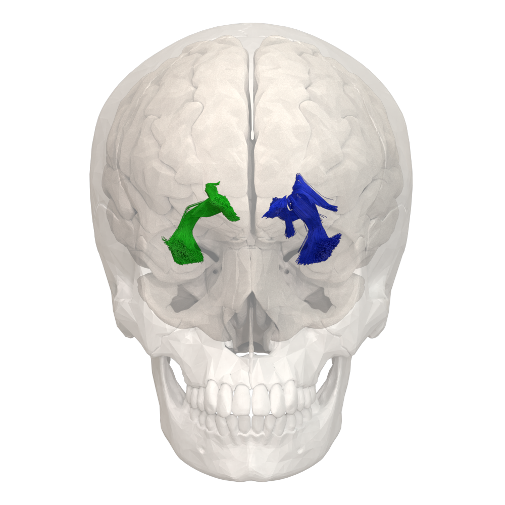
| 13:23, 8 May 2021 | Tractography - Uncinate fasciculus - anterior view.png (file) |  |
674 KB | == {{int:filedesc}} == {{Information |Description={{en|1=Tractography of the uncinate fasciculus. 3D Tractography data (by Fang-Cheng Yeh et al.) is superimposed on the skull and the brain from BodyParts3D. Rendering was done by Blender. (File conversion method). {{Legend|Blue|Blue: Left uncinate fasciculus}} {{Legend|Lime|Green: Right uncinate fasciculus}} }} |Source=Tractography data: http://brain.labsolver.org/diffusion-mri-tem... |
| 10:12, 8 May 2021 | Tractography - Uncinate fasciculus - animation 1.gif (file) |  |
10.2 MB | == {{int:filedesc}} == {{Information |Description={{en|1=Tractography of the uncinate fasciculus. Animation. 3D Tractography data (by Fang-Cheng Yeh et al.) is superimposed on the skull and the brain from BodyParts3D. Rendering was done by Blender. (File conversion method). {{Legend|Blue|Blue: Left uncinate fasciculus}} {{Legend|Green|Green: Right uncinate fasciculus}} }} |Source=Tractography data: http://brain.labsolver.org/diffu... |
| 15:35, 5 May 2021 | Tractography - Dentatothalamic tract - posterior view.png (file) |  |
685 KB | == {{int:filedesc}} == {{Information |Description={{en|1=Tractography of dentatothalamic tract. 3D Tractography data (by Fang-Cheng Yeh et al.) is superimposed on the skull and the brain from BodyParts3D. Rendering was done by Blender. (File conversion method). {{Legend|Blue|Blue: Right dentatothalamic tract<sup>*1</sup>}} {{Legend|Red|Red: Left dentatothalamic tract<sup>*1</sup>}} <sup>*1</sup> This left/right classification or nomenclature of the tracts... |
| 14:13, 5 May 2021 | Tractography - Dentatothalamic tract - animation 3.gif (file) |  |
7.15 MB | c:User:Rillke/bigChunkedUpload.js: == {{int:filedesc}} == {{Information |Description={{en|1=Tractography of dentatothalamic tract. Animation. Rendering was done by Blender. (File conversion method). {{Legend|Blue|Blue: Right dentatothalamic tract<sup>*1</sup>}} {{Legend|Red|Red: Left dentatothalamic tract<sup>*1</sup>}} <sup>*1</sup> This left/right classification or nomenclature of the tracts is by the HCP Tractography Atlas (Fang-Cheng Yeh et al., 20... |
| 13:42, 5 May 2021 | Tractography - Dentatothalamic tract - animation 2.gif (file) |  |
10.18 MB | c:User:Rillke/bigChunkedUpload.js: == {{int:filedesc}} == {{Information |Description={{en|1=Tractography of dentatothalamic tract. Animation. 3D Tractography data (by Fang-Cheng Yeh et al.) is superimposed on the skull and the brain from BodyParts3D. Rendering was done by Blender. (File conversion method). {{Legend|Blue|Blue: Right dentatothalamic tract<sup>*1</sup>}} {{Legend|Red|Red: Left dentatothalamic tract<sup>*1</sup>}} <sup>*1</sup> This left/r... |
| 11:31, 5 May 2021 | Tractography - Dentatothalamic tract - animation 1.gif (file) |  |
26.19 MB | c:User:Rillke/bigChunkedUpload.js: |
| 16:41, 3 May 2021 | Tractography - Cingulum - animation 3.gif (file) |  |
5.69 MB | == {{int:filedesc}} == {{Information |Description={{en|1=Tractography of cingulum. Anterior view. 3D Tractography data (by Fang-Cheng Yeh et al.) superimposed on the skull and brain (from BodyParts3D). Rendered by Blender.}} |Source=Tractography data: http://brain.labsolver.org/ Skull and brain data: http://lifesciencedb.jp/bp3d/ |Date=2021-04-30 |Author=Tractography data: Yeh, F. C., Panesar, S., Fernandes, D., Meola, A., Yoshino, M., Fernandez-Miranda, J. C., ... & Verstynen, T. (2018). Pop... |
| 16:01, 3 May 2021 | Tractography - Cingulum - animation 2.gif (file) |  |
8.55 MB | == {{int:filedesc}} == {{Information |Description={{en|1=Tractography of cingulum. Anterior view. 3D Tractography data (by Fang-Cheng Yeh et al.) superimposed on the skull and brain (from BodyParts3D). Rendered by Blender.}} |Source=Tractography data: http://brain.labsolver.org/ Skull and brain data: http://lifesciencedb.jp/bp3d/ |Date=2021-04-30 |Author=Tractography data: Yeh, F. C., Panesar, S., Fernandes, D., Meola, A., Yoshino, M., Fernandez-Miranda, J. C., ... & Verstynen, T. (2018). Pop... |
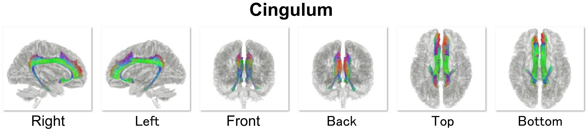
| 14:38, 3 May 2021 | Population-averaged Human tractography - Cingulum.png (file) | 428 KB | ||
| 06:00, 2 May 2021 | Different ways to subdivide the cingulum bundle.png (file) |  |
1.64 MB | {{Information |Description={{en|1=Cingulum bundle reconstructions based on diffusion MRI tractography. Images show the left cingulum for one healthy individual displayed on a T1-weighted image. A) The cingulum reconstructed as a single, continuous bundle (green), B) Dorsal (blue) and ventral (yellow) cingulum subdivisions (e.g., Budisavljevic et al., 2015). C) Subgenual (red), retrosplenial (orange), and parahippocampal (yellow) cingulum subdivisions (Jones et al., 2013). D) Proposed subdivis... |
| 04:46, 2 May 2021 | Connections of the cingulum bundle in macaque monkey.png (file) |  |
1.72 MB | {{Information |Description={{en|1=Schematic of macaque monkey brain showing connections that provide sagittal fibres to the cingulum bundle. (Note cingulate projections that cross the bundle, e.g., to the anterior thalamic nuclei, are not depicted). The colours help distinguish the multiple pathways. While it is most likely that additional subcortical projections join the cingulum, explicit descriptions are often lacking. Abbreviations: ACC, anterior cingulate cortex; ATN, anterior thalamic n... |
| 04:21, 2 May 2021 | Connections of the cingulum bundle in rat.png (file) |  |
1.47 MB | {{Information |Description={{en|1=Schematic of rat brain showing connections that provide sagittal fibres to the cingulum bundle. (Note cingulate projections that cross the bundle, e.g., to the anterior thalamic nuclei, are not depicted). The colours help distinguish the multiple pathways. Abbreviations: ACC, anterior cingulate cortex; ATN, anterior thalamic nuclei; CC, corpus callosum; DB, diagonal band; HPC, hippocampus, including subiculum; LC, locus coeruleus; LD laterodorsal thalamic nuc... |
| 02:21, 2 May 2021 | Five potential subcomponents of cingulum2.png (file) |  |
463 KB | {{Information |Description={{en|1= Spatial Relationship of the subcomponents of the cingulum bundle (CB). (A) A complete schematic map of the five segments. (B) Depicting five segments of CB, T1 sagittal view. The CB-I snugly curves around the genu of the CC and spreads into the orbital-frontal cortex as the lowest subdivision. For the projective scope in the medial aspect of the SFG, the CB-III is obviously larger than that of the CB-II. The body section of the CB-II extends along the bottom... |
| 02:17, 2 May 2021 | Five potential subcomponents of cingulum.png (file) |  |
277 KB | {{Information |Description={{en|1= In vivo fiber tractography of the cingulum bundle (CB) and five potential subcomponents in the CB. CB-I, red color; CB-II, green color; CB-III, yellow color; CB-IV, purple color; CB-V, blue color.}} |Source=Wu Y, Sun D, Wang Y, Wang Y and Ou S (2016) Segmentation of the Cingulum Bundle in the Human Brain: A New Perspective Based on DSI Tractography and Fiber Dissection Study. Front. Neuroanat. 10:84. doi: https://doi.org/10.3389/fnana.2016.00084 |Date=Publis... |
| 13:33, 1 May 2021 | Tractography - Cingulum - superior view.png (file) |  |
1.74 MB | == {{int:filedesc}} == {{Information |Description={{en|1=Tractography of cingulum. Superior view. 3D Tractography data (by Fang-Cheng Yeh et al.) superimposed on the skull and brain (from BodyParts3D). Rendered by Blender.}} |Source=Tractography data: http://brain.labsolver.org/ Skull and brain data: http://lifesciencedb.jp/bp3d/ |Date=2021-04-30 |Author=Tractography data: Yeh, F. C., Panesar, S., Fernandes, D., Meola, A., Yoshino, M., Fernandez-Miranda, J. C., ... & Verstynen, T. (2018). Pop... |
| 13:32, 1 May 2021 | Tractography - Cingulum - anterior view.png (file) |  |
1.64 MB | == {{int:filedesc}} == {{Information |Description={{en|1=Tractography of cingulum. Anterior view. 3D Tractography data (by Fang-Cheng Yeh et al.) superimposed on the skull and brain (from BodyParts3D). Rendered by Blender.}} |Source=Tractography data: http://brain.labsolver.org/ Skull and brain data: http://lifesciencedb.jp/bp3d/ |Date=2021-04-30 |Author=Tractography data: Yeh, F. C., Panesar, S., Fernandes, D., Meola, A., Yoshino, M., Fernandez-Miranda, J. C., ... & Verstynen, T. (2018). Pop... |
| 13:29, 1 May 2021 | Tractography - Cingulum - lateral view.png (file) |  |
2.02 MB | == {{int:filedesc}} == {{Information |Description={{en|1=Tractography of cingulum. Lateral view. 3D Tractography data (by Fang-Cheng Yeh et al.) superimposed on the skull and brain (from BodyParts3D). Rendered by Blender.}} |Source=Tractography data: http://brain.labsolver.org/ Skull and brain data: http://lifesciencedb.jp/bp3d/ |Date=2021-04-30 |Author=Tractography data: Yeh, F. C., Panesar, S., Fernandes, D., Meola, A., Yoshino, M., Fernandez-Miranda, J. C., ... & Verstynen, T. (2018). Popu... |
| 01:12, 1 May 2021 | Tractography - Cingulum - animation.gif (file) |  |
19.47 MB |
.png)
.png)
.png)
.png)
.png)
.png)